Microstructural analysis of P3D Scaffolds

New peer-reviewed article: Microstructural analysis of P3D Scaffolds using X-ray microcomputed tomography
Investigating bioceramic scaffolds with micro-CT
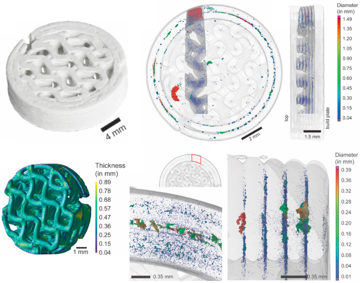
In a recent study, microcomputed tomography (micro-CT) was utilized to investigate the microstructural parameters of beta-tricalcium phosphate-based bone-mimicking scaffolds. Here, high and low-resolution microCT was used to quantitatively evaluate the 3D-printed, bone-mimicking constructs. The constructs are the surface area, construct volume, number of pores, pore size and distribution, wall thickness, delamination, internal porosity, and internal structure. In addition to establishing these parameters, this article underlines the value of microCT as a tool in the quality management of bioceramic implants.
Link to full article: Senck, S. et al. (2024) Ceramic additive manufacturing and microstructural analysis of tricalcium phosphate implants using X-ray microcomputed tomography. Open Ceramics. https://doi.org/10.1016/j.oceram.2024.100628

Visualization by first author, Sascha Senck, PhD, University of Applied Sciences Upper Austria.
What are the P3D Scaffolds?
Bioceramic 3D scaffolds that mimic the physical properties of bone – with no batch-to-batch variance.
The 3D printed structures replicate the architecture and complexity of calcified bone tissue. This let you create more relevant and reliable tissue- and disease models that capture the complex interplay between various cells.
The 3D scaffolds can also be used to test new therapies in lifelike structures wherein drug perfusion is uneven and where bacteria and cancer cells may hide in pores. This allows for more realistic testing that is more likely to yield reliable results when subsequently translated.
You get a biocompatible system of natural materials and customized structures that let you create predictive research models of human physiology and pathology.


