Contact us here!
Contact us for questions, general inquiries, or to request a quote.
By submitting this form, I consent to the processing of my personal data as explained in Ossiform's Privacy Policy.
Contact us for questions, general inquiries, or to request a quote.
By submitting this form, I consent to the processing of my personal data as explained in Ossiform's Privacy Policy.
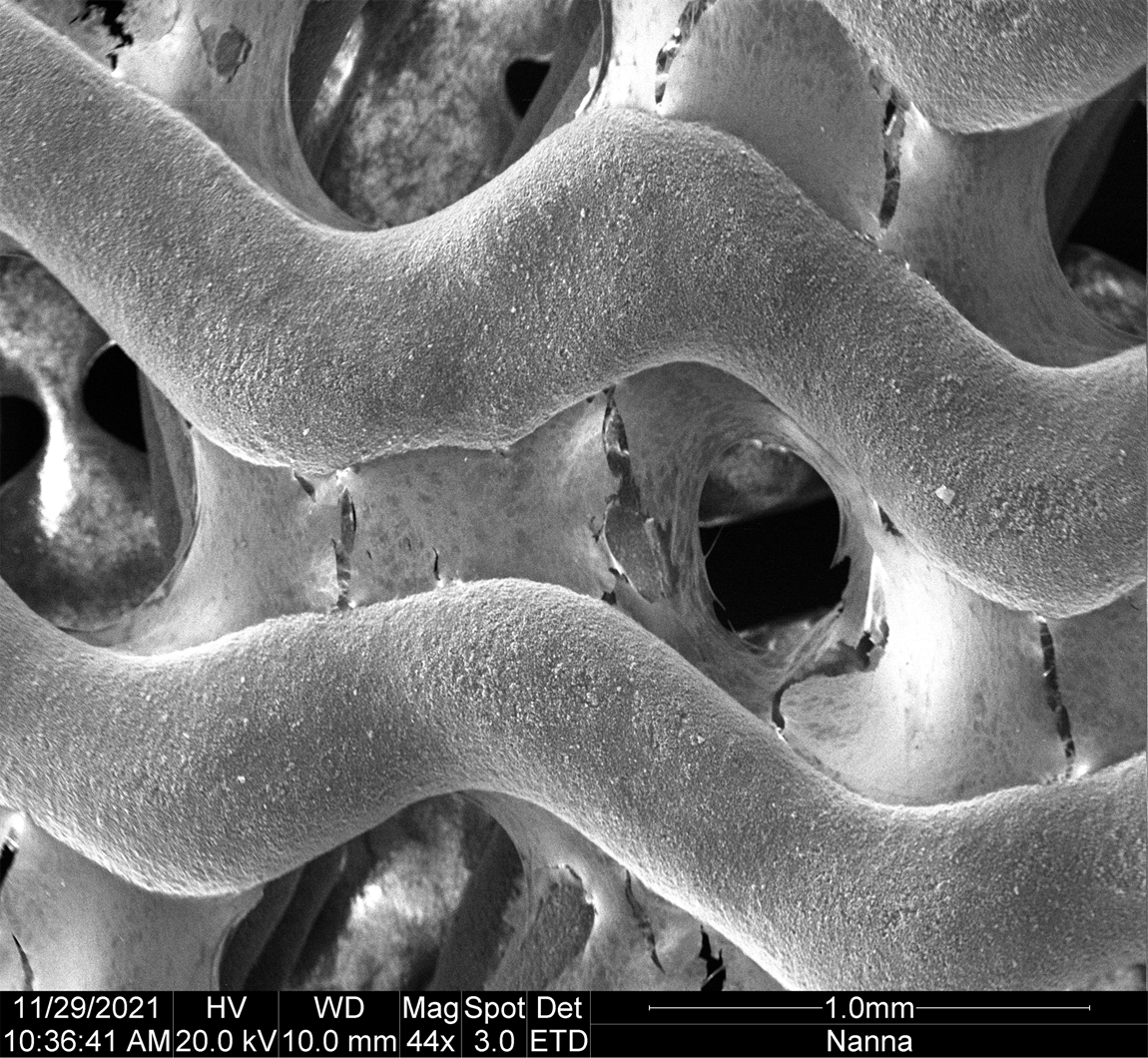
Earlier this week, our in-house scientist, Nanna, used SEM imaging to evaluate if osteoblasts cultured on the P3D Scaffolds are producing collagen. This led to several intriguing images that show a large amount of collagen produced.
The picture shown in this weeks “News from the Lab” illustrates how the P3D Scaffolds allow osteoblasts to produce an extra cellular matrix that spans throughout the scaffold in an in vivo like manner.
If you want to learn more about culturing of cells on our P3D scaffolds, please visit our resources page.

Bioceramic 3D scaffolds that mimic the physical properties of bone – with no batch-to-batch variance.
The 3D printed structures replicate the architecture and complexity of calcified bone tissue. This lets you create more relevant and reliable tissue- and disease models that capture the complex interplay between various cells.
The 3D scaffolds can also be used to test new therapies in lifelike structures. This allows for a more realistic testing that is more likely to yield reliable results when subsequently translated.
You get a biocompatible system of natural materials and customized structures. This lets you create predictive research models of human physiology and pathology.