Contact us here!
Contact us for questions, general inquiries, or to request a quote.
By submitting this form, I consent to the processing of my personal data as explained in Ossiform's Privacy Policy.
Contact us for questions, general inquiries, or to request a quote.
By submitting this form, I consent to the processing of my personal data as explained in Ossiform's Privacy Policy.

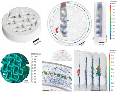
Visualization by first author, Sascha Senck, PhD, University of Applied Sciences Upper Austria.
In a study from 2024, microcomputed tomography (micro-CT) was utilized to investigate the microstructural parameters of 3D printed β-tricalcium phosphate-based bone-mimicking scaffolds. Micro-CT is a 3D imaging technique that utilizes X-rays to visualize internal structures of objects at a high resolution in a non-destructive manner.
Verifying the internal microstructure of an implant can be done in numerous ways that can be both destructive and non-destructive to the 3D printed constructs. Non-destructive methods are useful in various industries, including the bone implant industry, since it can be employed in the quality management process to ensure that the implant lives up to the specified requirements. Industrial micro-CT is one of the most promising non-destructive methods for the in-depth examination of constructs such as implants due to its high resolution and rapidness.
In the study, high- and low-resolution micro-CT was used to quantitatively evaluate the 3D printed, bone-mimicking constructs, P3D Scaffolds from Ossiform®. These scaffolds were designed with an outer dense rim and internal gyroid lattice structure. The examined parameters were: surface area, construct volume, number of pores, pore size and distribution, wall thickness, delamination, internal porosity, and internal structure. Expectedly, the quantitative results of most of these parameters largely depended on the chosen scanning resolution. For example, the higher scanning resolution detected a larger number of smaller pores than the low-resolution scan. Resultingly, mean pore size was significantly different between the scans due to greater detection of smaller objects for the high-resolution scan.
If the quantitative result of a scan is that dependent on the resolution, then why not always scan at the highest possible resolution? This is because the resolution of the scan is affected by specimen size, meaning that scanning of larger objects will result in lower spatial resolutions. Higher resolution results with greater detectability can be obtained in large specimens by only scanning a chosen region of interest (ROI) at a time. This in turn significantly increases the time it takes to perform the full scan as well as introduces the risk of scanning artefacts. Alternatively, a representative ROI for the whole specimen can be chosen and analyzed, but at the expense of risking missing important information outside of the ROI.
Effectively visualizing the internal structure of implants, including wall thickness, pore size and pore distribution, is crucial in the design evaluation of the construct. This ensures that the mechanical strength and stiffness is balanced with the capabilities of supporting osseointegration with the native bone.
Learn more about the findings and how micro-CT can be a great tool in the orthopedic bone implant industry. Read the full article here:
Senck, S. et al. (2024) Ceramic additive manufacturing and microstructural analysis of tricalcium phosphate implants using X-ray microcomputed tomography. Open Ceramics. https://doi.org/10.1016/j.oceram.2024.100628
The P3D Scaffolds are bioceramic 3D printed scaffolds made from β-TCP, which mimic the physical properties of bone – with no batch-to-batch variance. The internal structures replicate the architecture and complexity of calcified bone tissue. This lets you create more relevant and reliable tissue- and disease models that capture the complex interplay between various cells.
The scaffolds can also be used to test new therapies in lifelike structures wherein drug perfusion is uneven and where bacteria and cancer cells may hide in pores. This allows for more realistic testing that is more likely to yield reliable results when subsequently translated.
You get a biocompatible system of natural materials and customized structures that let you create predictive research models of human physiology and pathology.