Contact us here!
Contact us for questions, general inquiries, or to request a quote.
By submitting this form, I consent to the processing of my personal data as explained in Ossiform's Privacy Policy.
Contact us for questions, general inquiries, or to request a quote.
By submitting this form, I consent to the processing of my personal data as explained in Ossiform's Privacy Policy.

Our bones have always been a central part of our existence as humans. More than a million years ago, early hominins were already making use of bone as raw material for the shaping of tools, demonstrating both practicality and ingenuity. With the emergence of civilization, the curiosity and human need to understand the world also flourished. Ancient Greek physicians and philosophers dissected animals, and occasionally humans, in the quest to understand the human body and anatomy. During the Renaissance, further refinement of anatomical understanding took place, most famously through Leonardo da Vinci’s detailed drawings, which bridged art and scientific observation. Shortly after, the foundations of modern bone biology were shaped with early description of bone growth, macroscopic features, and microscopic structures such as Haversian canals. With the development of microscopy, entirely new dimensions opened as researchers were suddenly able to observe our bone architecture on a finer scale than ever before. Yet even today, numerous questions about the very framework of our bodies are still largely unanswered and remain an active frontier of scientific discovery.
Today, bone research is at the cusp of advancement. While traditional 2D cell culturing experiments and investigations in animals have provided great insights into the cellular and molecular mechanisms of bones over the years, their lack of physiological relevance to human biology is still present.
Although 2D cell culturing provides ease of use and is cost and time efficient, they do not provide the cells with a physiologically relevant 3D environment. Rather more, they only allow the cells to grow in a monolayer on a rigid surface without the natural substrate, concentration gradients and stiffness of the native extracellular matrix. This can result in alterations to the cellular morphology and behavior, limited cell-to-cell and cell-to-matrix interactions, poor predictivity for drug testing and lack of translational results. Furthermore, 2D cell cultures are not optimized for investigating complex cellular interactions such as the ones in co-cultures, thereby limiting the degree of model complexity.
On the other end of the spectrum of pre-clinical research models lies animal models. They provide physiological context to the setup, where cellular diversity, native extracellular matrix, and the immune system are represented. However, they are time-consuming and costly, introduce significant biological variation, and show poor translation to human biology as well as poor drug predictability. Additionally, researchers, regulatory authorities, and the industry are increasingly focused on limiting the use of animals in research for ethical reasons.
Due to the limitations of the traditional research models, the advancement of more complex setups such as 3D scaffolds, organoids and spheroids, organ-on-a-chip technologies, and bioreactors have been rapid over the last few decades. These combine the relative simplicity, controllability, ease of use, and cost efficiency of the cell culture setup with the increased complexity and improved physiological relevance of the animal models without their drawbacks. These advances highlight the growing need for biomimetic, controllable, and ethically responsible research platforms. These are needs that Ossiform® aims to address with its P3D Scaffolds.
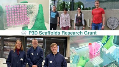
In our continuous efforts to support the advancement of current research, Ossiform® initiated a grant project where we would supply chosen research groups with the necessary P3D Scaffolds for their project of choice. Numerous applicants showed interest and sent in their project descriptions. Researchers at the University of Oxford and the University of Southern Denmark were the recipients of the Ossiform® grant and the P3D Scaffolds. The goal of the grant project is to help researchers simulate a bone-relevant in vivo environment.
A plethora of publications have elucidated detailed molecular- and biophysical mechanisms of membrane transporters. However, much less has been published about how cells effectively maneuver the complex 3D environments of living tissues. A research team from the University of Southern Denmark, led by Professor Carsten Uhd Nielsen, aims to study the function of membrane transporters. They will be using Ossiform’s P3D Scaffolds as an appropriate platform. With this technology, the group aspires to simulate a dynamic in vivo-like environment to facilitate pharmaceutical applications related to membrane transporters.
Currently, the prevention of prostate cancer bone metastasis is halted by palliative treatments and limited preclinical secondary bone cancer models. Consequently, no curative treatment exists for this cancer stage. However, researchers at the University of Oxford, led by Jia-Ling Ruan, aspire to advance appropriate therapies. They are integrating Ossiform’s P3D Scaffolds in their 3D tumor/organoid cultures. The aim is to develop an effective in vitro bone metastasis model. Utilizing FLASH radiotherapy, the researchers can effectively study the effects of radiation responses of normal bone tissue, tumor tissue, and the abscopal effects of secondary bone metastasis, respectively.
The P3D Scaffolds from Ossiform® are ceramic scaffolds made entirely of β-tricalcium phosphate – a mineral that effectively mimics the calcified part of our bones and is recognized as such by bone cells such as osteoblasts. This allows researchers to grow their bone-derived cells in a clinically and physiologically relevant environment without the use of any animal-derived materials and with no batch-to-batch variability. The scaffolds’ interconnected porosity allows cells to migrate, organize, and form tissue‑like structures in three dimensions.
The P3D Scaffolds are easy to use, stable in culture and can be combined with traditional laboratory techniques with little to no alterations to the existing protocols. They allow culturing complex tissue and disease models through co-culturing and can be used for drug testing with repeated dosing.
Learn more about the P3D Scaffolds here and find our protocols here.