Contact us here!
Contact us for questions, general inquiries, or to request a quote.
By submitting this form, I consent to the processing of my personal data as explained in Ossiform's Privacy Policy.

Contact us for questions, general inquiries, or to request a quote.
By submitting this form, I consent to the processing of my personal data as explained in Ossiform's Privacy Policy.
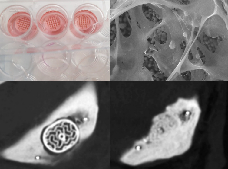
Understanding human disease and developing effective therapies depend heavily on the experimental models used in research. For decades, scientists have relied on simplified in vitro systems and animal models to study complex biological processes, but these approaches often fail to fully capture human physiology. For in vitro models, important aspects of tissue architecture, cell–cell interactions, and microenvironmental cues are frequently lost, limiting the predictive value of traditional models. On the other hand, the use of animal models entails greater biological variability and lack predictive accuracy due to fundamental differences compared to human physiology and pathology.
In recent years, advances in biomaterials, 3D printing, and cell culture technologies have enabled the development of more physiologically relevant experimental systems. Among these, 3D cell culture has emerged as a powerful approach to better replicate native tissue environments, offering new opportunities to study disease mechanisms and evaluate therapeutic strategies in a context that more closely resembles the human body.
With growing recognition of their limitations, 3D cell culture has gained increasing attention as a way to bridge the gap between simplified in vitro systems and complex human biology. In fact, in vivo animal testing is currently required to document the safety and efficacy of treatments in a living organism. However, animal models are time, labor, and resource intensive and are not always a reliable way to predict how drug treatments will affect humans. Beyond improving biological relevance, 3D cell culture systems also align with the growing emphasis on the 3Rs (Replacement, Reduction, and Refinement) in animal research as well as offering an opportunity to generate meaningful data earlier in the drug development pipeline. Meanwhile, the traditional two-dimensional cell culture models do not accurately mimic the cellular environments of human tissues or physiology. Namely, because the cells in our body do not grow flat in a monolayer with bulk concentrations of nutrients, cytokines and cell signaling substances. While the limitations of 2D cultures and animal models are well documented, their continued use highlights the lack of sufficiently predictive in vitro alternatives.
3D cell culture systems, on the other hand, are more physiologically relevant and reliable research models as they maintain the cell-to-cell interaction as well as the cell-to-matrix interaction without the variability of animal models. They can be grown on scaffolds that offer an environment which allow the cells to follow their own genetic instructions to self-organize and form 3D structures like they would inside the body. As a result, 3D models are increasingly used to inform earlier decision‑making in research workflows, where predictive accuracy is critical. Reducing the use of in vivo animal testing, owing to the relevant and predictive data from 3D cellular models, may consequently reduce the costs and time needed to get new human therapeutics into the clinic. This shift also aligns with growing ethical, economic, and regulatory pressure to reduce reliance on animal testing, as well as the need to generate meaningful data earlier in the drug development process.
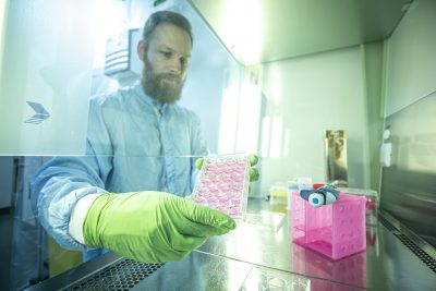
The value of 3D cell culture becomes particularly evident when studying diseases that arise within complex tissue environments, such as bone cancer. Bone tissue is characterized by a highly specialized extracellular matrix, distinct mechanical properties, and tightly regulated cell–cell and cell–matrix interactions. Replicating these features in experimental models has long been a challenge, and as a result, robust and predictive in vitro systems of primary bone cancer have been limited.
In cancer drug screening, conventional 2D monolayer cell cultures fail to recapitulate the histoarchitecture of bone tumors as well as key aspects of tumor heterogeneity and mechanical signaling. 3D cell culture systems, such as the P3D Scaffolds, provide a more physiologically relevant environment by mimicking both the composition and stiffness of native bone tissue. This enables cancer cells to grow in three dimensions, interact dynamically with the surrounding matrix, and establish gradients of oxygen, nutrients, and metabolites similar to those found in solid tumors. These features are particularly important in bone cancer, where mechanical cues and matrix interactions play a central role in tumor progression and treatment response. Importantly, these are also central to regenerative medicine, where cell fate, migration, and tissue remodeling are strongly influenced by the physical and biochemical properties of the extracellular matrix.
By recreating key aspects of the bone microenvironment, 3D cell culture systems allow for realistic assessments of drug efficacy and resistance in cancer patients. Uneven drug penetration, variable cell proliferation rates, and the presence of genetically and phenotypically diverse cell populations can all be captured more effectively in 3D systems. This increased biological relevance enables researchers to identify promising therapeutic candidates earlier, while also revealing ineffective approaches sooner than is typically possible with 2D cultures. Furthermore, researchers are also able to study how malignant cells interact with bone‑forming cells, degrade or remodel the surrounding matrix, and influence tissue regeneration. These insights are relevant for understanding tumor‑induced bone destruction as well as for developing regenerative strategies aimed at restoring bone structure and function following disease or treatment.
Ultimately, these advances improve the efficiency and translational value of preclinical research, supporting faster and more informed decision‑making in the development of new therapies.
Translating these insights into practical research tools requires experimental systems that faithfully replicate the structural and material properties of bone tissue in a controlled laboratory setting.
Ossiform’s P3D Scaffolds are lifelike bone environments made from β-tricalcium phosphate using a proprietary 3D printing process. The unique structures are 3D printed with porosities to mimic the complex porous structure observed in human bone tissues. The result is a cell growth support structure and tissue model that yields predictive research models of human biology, that allows researchers to perform relevant and accurate cellular environment investigations.
While the P3D Scaffolds are not certified for use in humans, they can be used for a wide range of pre-clinical in vitro and in vivo research studies. As 3D cell culture technologies continue to mature, lifelike tissue models are expected to play an increasingly central role in biomedical research. By bridging the gap between simplified in vitro systems and complex in vivo biology, 3D models have the potential to accelerate discovery, improve translational success, and ultimately support the development of more effective therapies in both disease modeling and regenerative medicine.
Find the peer-reviewed scientific publications using P3D Scaffolds here.
