Contact us here!
Contact us for questions, general inquiries, or to request a quote.
By submitting this form, I consent to the processing of my personal data as explained in Ossiform's Privacy Policy.
Contact us for questions, general inquiries, or to request a quote.
By submitting this form, I consent to the processing of my personal data as explained in Ossiform's Privacy Policy.
Ossiform is pleased to share that a new peer-reviewed research article featuring its P3D Scaffolds has been published in ACS Omega.
The P3D scaffolds are part of Ossiform Research Line and for Research Use Only.
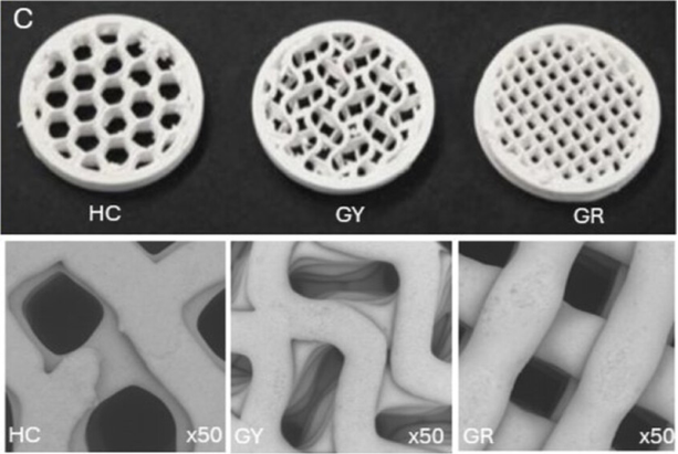
For decades, the architectural effects on mechanical strength and biological outcomes of bone substitutes have been investigated. In this study, seven scaffold systems were compared: PCL nanofibrous membrane made by Direct Electrospin Writing (DEW), 3D printed PCL with different infill architectures (grid, gyroid or honeycomb), and Ossiform’s 3D printed, beta-tricalcium phosphate (b-TCP) P3D Scaffolds with the same three infill structures (grid, gyroid or honeycomb).
Thapa S., et al. (2026). Macro images of 3D-printed β-TCP scaffolds sourced from Ossiform in honeycomb (HC), gyroid (GY), and grid (GR) configurations, respectively. SEM micrographs showing characteristic fiber/pore architectures across scaffold types under 50× magnification.
Using Scanning Electron Microscopy (SEM) imaging, it was that the P3D Scaffolds have a granular surface microstructure, which is uniformly distributed across the scaffold surface and contributes to significant surface roughness and microporosity. Both the surface roughness and microporosities are favorable for cellular attachment and proliferation, and consistent with osteoconductive surface characteristics reported for calcium phosphate ceramics, which mimic the hierarchical topography of natural bones. This is in contrast to the microstructural surface of PCL, which is smooth and therefore does not provide much support for cellular adhesion.
During mechanical tests, the P3D Scaffolds demonstrated the highest level of stiffness of the tested scaffolds and exhibited the expected brittleness inherent to the material. Under uniaxial tensile testing, the β‑TCP scaffolds showed a high elastic modulus but underwent abrupt failure at stresses around 70–80 MPa, reflecting the brittle nature of the material.
Furthermore, biological outcomes of scaffold material and architecture were investigated using direct culturing of osteosarcoma cells (MG63) for 7 days, where cellular morphology, proliferation, and viability were examined.
On the ceramic P3D Scaffolds from Ossiform, early effects on cell morphology and attachment were dependent on scaffold architecture. The gyroid scaffolds showed superior early attachment and elongated morphology compared to the grid and honeycomb designs that had cells with less spread and more rounded morphology. All three b-TCP scaffold designs showed supported spreading of cells over time, but the gyroid scaffolds consistently showed the highest degree of cell elongation compared to the two others, indicating continuous elongation and guidance cues along the curved surface of the scaffold fibers. Like it was also observed with the PCL scaffolds, the grid and honeycomb P3D Scaffolds promoted more isotropic spreading of cells, which could be attributed to their flat surfaces and repeating geometry. This trend persisted until day 7, where the gyroid scaffolds also showed the highest degree of cellular bridging. Similar trends favoring the gyroid P3D Scaffold from Ossiform were observed by the cellular viability assay. These observations suggest that the gyroid structure is superior in terms of cellular support for this cell line, which may be attributed to their curved design that may enable effective mechanotransduction, as well as to their large surface area, as inferred by the authors.
While the cells grown on the PCL-DEW scaffolds initially showed rounded morphology and limited spreading, they experienced the highest degree of viability and proliferation of all the scaffolds on day 7. Furthermore, they demonstrated increased elongation and spreading morphology that aligned with the nanofiber orientation over time. On the 3D printed PCL scaffolds, cellular morphology in the initial phase of cell culturing was influenced by scaffold architecture with more attached and elongated cells on the grid scaffolds compared to the honeycomb and gyroid structures. Spreading and elongation was dramatically enhanced at day 7 on the grid and gyroid PCL scaffolds, where bridging connections between scaffold layers were observed. On the grid PCL scaffolds, there were features suggestive of early fiber degradation and deposition of extracellular matrix-like material. On the PCL honeycomb scaffolds, cellular morphology appeared more spread and random without the strong uniaxial alignment as seen on the grid scaffold on day 7. These morphological findings were supported by the viability assay, where the highest proliferation was observed on the gyroid PCL scaffold. Furthermore, the gyroid structure of the PCL scaffolds demonstrated superior elongation, which formed well-spread morphologies that effectively adhered to the curved surfaces of the scaffold, suggesting that the complex microenvironment facilitated multidirectional migration. Similar effects were less evident on the grid and honeycomb PCL structures, where the flat nature of the geometry limited lateral spreading, decreased cellular elongation, and probably resulted in the decreased cellular viability compared to the gyroid architecture.
Overall, the study concludes that different choices of material and design achieve different goals. Where PCL scaffolds benefit from their elasticity, ductility, and energy absorption, β-TCP exhibits a higher elastic modulus and fracture stress under tensile loading but is more brittle. Furthermore, the authors suggest that β-TCP scaffolds benefit from their surface chemistry for osteogenesis, while PCL scaffolds benefit from their mechanical elasticity and ductility in general, and nanoscale topography for the PCL-DEW scaffold. Regarding the mechanical strength provided by the scaffold architecture, grid showed the highest stiffness, honeycomb had the most balanced load-bearing, and gyroid showcased the most uniform distribution of strain. Lastly, cellular morphology and proliferation data showed favorable results toward the gyroid architecture with increased elongation, spreading, bridging, and viability compared to the grid and honeycomb structures. However, it is important to note that these results are derived from short-term cell cultures and mechanical tests performed in non-hydrated settings, and no in vivo data is included, as stated by the authors.
Read the full article here: Scaffold Material–Architecture Design Rules Linking Mechanics and Early Osteogenesis in PCL/β-TCP Grid, Honeycomb, and Gyroid Lattices | ACS Omega
Disclaimer: the paper states that the P3D Scaffolds from Ossiform are produced through freeze-drying. This is incorrect as the P3D Scaffolds are produced through 3D printing following sintering at high temperatures.