Contact us here!
Contact us for questions, general inquiries, or to request a quote.
By submitting this form, I consent to the processing of my personal data as explained in Ossiform's Privacy Policy.
Contact us for questions, general inquiries, or to request a quote.
By submitting this form, I consent to the processing of my personal data as explained in Ossiform's Privacy Policy.
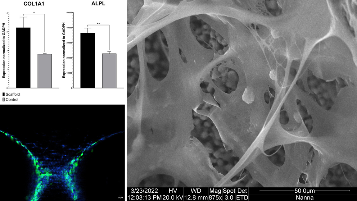
Our latest studies have shown that the P3D Scaffolds have great osteogenic properties. The studies also show that osteoblasts maintain their genotype and morphological phenotype. We have had the osteoblasts cultured on the bone-mimicking bioceramic scaffolds for 2-5 weeks and the results are clear.
Osteoblastic marker genes are upregulated in response to traditional cell culturing. Confocal imaging shows how the P3D scaffolds allow the osteoblasts to organize in a 3D-oriented manner. All whilst surrounded by their collagen-rich extracellular matrix.
Studying the osteoblasts and their extracellular matrix closer using scanning electron microscopy, you can appreciate how the osteoblasts retain their round morphology. Furthermore, you can see how the extracellular matrix is spanning widely across the scaffold, clearly indicative of the first steps in bone formation.

Bioceramic 3D scaffolds that mimic the physical properties of bone – with no batch-to-batch variance.
The 3D printed structures replicate the architecture and complexity of calcified bone tissue. This lets you create more relevant and reliable tissue- and disease models that capture the complex interplay between various cells.
The 3D scaffolds can also be used to test new therapies in lifelike structures. This allows for a more realistic testing that is more likely to yield reliable results when subsequently translated.
You get a biocompatible system of natural materials and customized structures. This lets you create predictive research models of human physiology and pathology.