Contact us here!
Contact us for questions, general inquiries, or to request a quote.
By submitting this form, I consent to the processing of my personal data as explained in Ossiform's Privacy Policy.

Contact us for questions, general inquiries, or to request a quote.
By submitting this form, I consent to the processing of my personal data as explained in Ossiform's Privacy Policy.
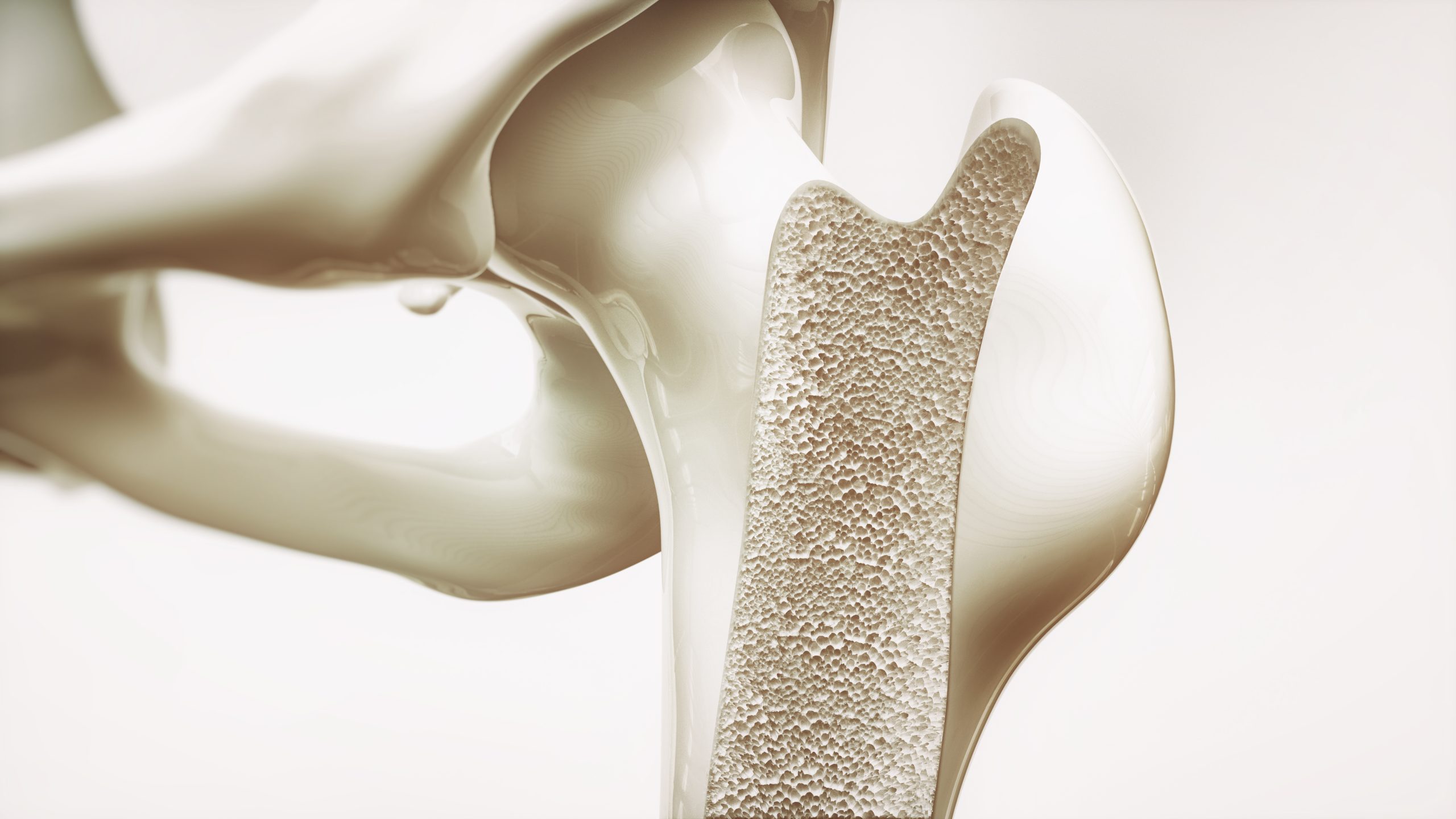
The human body is composed of multiple specialized tissue types each with unique properties tailored to support the diverse functions necessary to stay healthy. One such tissue is our bones, which most would characterize as static and rigid, calcified structures that form the skeleton, act as a scaffold for our soft tissues, and protect vital organs. However, what many don’t know is that the skeleton is a highly dynamic, specialized, and flexible tissue that not only remodels continuously but performs diverse functions affecting the whole body (Florencio-Silva et al., 2015).
Like other tissues and organs, the bones are a part of a complex and interconnected system with a delicate balance. Comprehensive insights into the physiology of our bones provides the basis for understanding what happens when the balances are tipped, homeostasis is disrupted, and pathophysiological states occur.
Overall, the bone tissue is compartmentalized into the bone matrix and the bone marrow, where the matrix is the hard, calcified part that most associates with the tissue.
The main components of the bone matrix are inorganic hydroxyapatite crystals, organic collagen, and non-collagenous proteins(Florencio-Silva et al., 2015; Viguet-Carrin et al., 2006). Hydroxyapatite is a calcium phosphate mineral, which,together with smaller amounts of other minerals and trace elements, constitutes 70% of our bone tissue. This inorganic part accounts for the inherenthardnessand stability of our bones but alone it would make the tissue brittle, unflexible, and easy to break.
By combining the elasticity of collagen with the hardness of the hydroxyapatite crystals, our bones become strong yet flexible, which ensure that our skeleton can withhold the stresses and compression that the skeleton is exposed to without breaking or losing its shape (Viguet-Carrin et al., 2006).
The bone marrow, on the other hand, is the soft and spongy tissue found inside the hollow center of most of our bones, which can be divided into red marrow and yellow marrow. The red marrow is primarily localized in large and highly vascularized bones, is an active tissue that serves as a stem cell niche and is the primary location for production of blood cells. In turn, the yellow marrow is primarily present in the long bones and is mostly composed of fat that functions as energy storage. During growth and ageing, the red marrow can turn into yellow marrow, but yellow marrow can also, under certain conditions, revert into red marrow to increase the blood cell production (Florencio-Silva et al., 2015).
Evidently, our skeleton is not merely a static tissue acting as a structural scaffold but consists of important minerals and cells important for our normal function and health. While important, the composition of our bones alone does not account for its properties but is further supported by the matrix turnover, density, and highly specialized architecture.
While the composition of the bones provides the foundation for their mechanical properties, the organization of the components are equally important. Bone tissue is arranged into highly specialized structures that maximize strength while minimizing weight, allowing the skeleton to support the body, facilitate movement, and protect internal organs (Florencio‑Silva et al., 2015; Seeman & Delmas, 2006).
Besides being divided into matrix and marrow, the bone tissue can also be divided into compact bone (cortical bone) and trabecular bone (cancellous or spongy bone). The cortical bone forms the dense outer shell of most bones, accounts for approximately 80% of the skeletal mass, and provides most of the vital mechanical strength of the tissue. This part of the bone is further organized into its fundamental structural units, called osteons or the Haversian system. Osteons consist of concentric layers of mineralized bone matrix, called lamellae, that are arranged around a central Haversian canal that in turn contains nerves and blood vessels. The osteons are interconnected by transverse channels that facilitates nutrient transport throughout the tissue and allows the compact bone to be metabolically active despite its density (Florencio-Silva et al., 2015).
In contrast, the cancellous bone is predominantly found at the ends of long bones and in the vertebrae. Here, it forms a porous, lattice-like structure with interconnected rods and plates known as trabeculae. This structure reduces the weight of the bones while maintaining the structural integrity since the trabeculae are strategically aligned along the lines of mechanical stress and thereby allowing it to distribute the mechanical loads. The spaces between the trabeculae are filled with the bone marrow, creating a close relationship between the skeletal and hematopoietic systems. Due to its large surface area and high metabolic activity, trabecular bone is the primary site of bone remodeling (Grabowski, 2009; Kenkre & Bassett, 2018).
Together, the organization of compact and trabecular bone allows the skeleton to achieve an optimal balance between strength, flexibility, and weight. Importantly, these structures are not static as they are continuously reshaped and adapted through the process of bone remodeling in response to mechanical loading, injury, ageing, and changes in physiological demand (Florencio‑Silva et al., 2015; Seeman & Delmas, 2006).
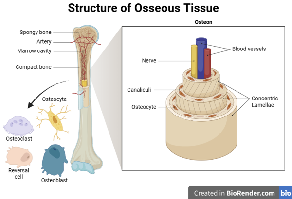
Figure 1: Illustration of osseous tissue and cell types in the bones. Created in BioRender.com.
Maintaining the intricate and dynamic architecture of the bones requires coordinated activity of several specialized cells. Besides the adipocytic cells and stem cells in the bone marrow niche, the calcified part of our bones contains a great variety of cells. These cells continuously monitor, remove, and rebuild bone tissue, ensuring that the skeleton remains structurally stable and capable of adapting to physiological demand and stress (Raggatt & Partridge, 2010). The cells include osteoblasts, osteoclasts, osteocytes, and reversal cells.
Osteoblasts are bone forming cells and arise from differentiation of osteogenic cells. The osteoblasts create new mineralized bone by producing the collagen-rich extracellular matrix and subsequently deposit calcium on the collagen fibers. Besides producing bone matrix, osteoblasts also regulate the activity of osteoclasts through secretion of signaling molecules such as Receptor Activator of Nuclear Factor-κB Ligand (RANKL) and osteoprotegerin (OPG), thereby playing an important role in coordinating the whole remodeling process (Raggatt & Partridge, 2010; Florencio-Silva et al., 2015).
Osteoclasts are the counter opposites of osteoblasts. They originate from the hematopoietic stem cell lineage in the bone marrow, and their key responsibility is to resorb old bone tissue through the production of proteolytic enzymes and secretion of hydrogen ions. This process releases skeletal minerals, which is vital for managing the extracellular calcium content, and it allows solid skeletal structures to be replaced by a hollower skeletal architecture with a better strength-to-weight ratio (Grabowski, 2009; Raggatt & Partridge, 2010; Kenkre & Bassett, 2018).
Osteocytes are terminally differentiated osteoblasts integrated in the bone matrix that sense the structural integrity and inflammation of bone. Osteocytes constitute more than 90% of all bone cells and form an extensive communication network through microscopic channels known as canaliculi. Through this network, osteocytes act as the primary mechanosensors of bone tissue, detecting mechanical loading and microdamage and coordinate the remodeling response accordingly (Raggatt & Partridge, 2010; Xiao et al., 2016; Arias et al., 2018).
Lastly, the reversal cells remove matrix debris and produce the coupling signal between resorption and formation of bone. Although less studied than osteoblasts and osteoclasts, reversal cells are increasingly recognized as important coordinators of remodeling. They help prepare the bone surface for subsequent bone formation and contribute to communication between the resorption and formation phases (Raggatt & Partridge, 2010; Delaisse et al., 2020).
Together, these cell types form a tightly coordinated biological system that allows bones to continuously adapt to changing mechanical and physiological demands (Raggatt & Partridge, 2010; Kenkre & Bassett, 2018).
Bone remodeling is a continuous process, where old, damaged, or mechanically compromised bone tissue is removed and replaced with new bone tissue. However, it not only preserves skeletal strength but also contributes to the regulation of mineral homeostasis by enabling the controlled release and storage of calcium and phosphate. To ensure proper skeletal maintenance, bone remodeling is tightly regulated by both local cellular signals and systemic hormones (Raggatt & Partridge, 2010; Kenkre & Bassett, 2018; Hadjidakis & Androulakis, 2006).
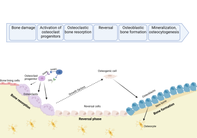
Figure 2: Illustration of the bone remodeling process. Created in BioRender.com.
The remodeling cycle begins when osteoclast precursors are recruited to a specific location on the bone surface and differentiate into active osteoclasts. These cells attach to the bone and create an isolated microenvironment in which they dissolve the mineralized matrix and degrade its organic components. Once the resorption phase is complete, the osteoclasts undergo programmed cell death and the exposed surface is prepared for bone formation by reversal cells (Raggatt & Partridge, 2010; Kenkre & Bassett, 2018).
The subsequent formation phase is initiated by signaling molecules released during bone resorption, including growth factors stored within the bone matrix. These signals recruit mesenchymal stem cells and early osteoblast progenitors to the remodeling site, where they differentiate into mature osteoblasts that produce a new unmineralized matrix consisting primarily of type I collagen and other extracellular matrix proteins. This matrix is subsequently mineralized through the deposition of hydroxyapatite crystals, restoring the strength and function of the tissue (Raggatt & Partridge, 2010; Arias et al., 2018; Hadjidakis & Androulakis, 2006).
Under healthy conditions, the amount of bone removed by osteoclasts is balanced by an equivalent amount of new bone formed by osteoblasts. This coupling between resorption and formation allows the skeleton to continuously adapt to mechanical loading, repair microdamage, and maintain optimal bone quality throughout life (Kenkre & Bassett, 2018; Arias et al., 2018).
Beyond their structural and mechanical functions, bones also play an important role in maintaining the body’s mineral balance and are increasingly recognized as an endocrine organ. In particular, the skeleton serves as the largest reservoir of calcium and phosphate in the body. Approximately 99% of the body’s calcium and 85% of its phosphate are stored within the mineralized bone matrix, making bone a critical regulator of mineral homeostasis (Grabowski, 2009; Raggatt & Partridge, 2010).
Maintaining stable calcium concentrations in the bloodstream is essential for many physiological processes, including muscle contraction, nerve signaling, blood clotting, and cellular communication. For this reason, calcium levels are tightly regulated through the coordinated actions of the skeleton, kidneys, intestines, and parathyroid glands.
When blood calcium levels decrease, the parathyroid glands release parathyroid hormone (PTH). PTH binds to receptors on osteoblastic cells, causing increased expression of RANKL and reduced expression of OPG. This shifts the remodeling balance toward osteoclast formation and activation, resulting in increased bone resorption and release of calcium and phosphate from the bone matrix into the bloodstream. Simultaneously, PTH acts on the kidneys to conserve calcium and stimulate the activation of vitamin D, which enhances calcium absorption from the intestines. Conversely, when calcium concentrations are elevated, the thyroid gland secretes calcitonin, which inhibits osteoclast activity and reduces bone resorption. Although the role of calcitonin in adult humans appears more limited than that of PTH, it contributes to the overall regulation of calcium homeostasis (Grabowski, 2009; Raggatt & Partridge, 2010).
In recent years, bones have also emerged as active endocrine organs capable of influencing distant tissues. Osteoblasts produce signaling molecules such as osteocalcin, which has been implicated in regulating glucose metabolism, energy expenditure, and reproductive function (Karsenty & Ferron, 2012). These discoveries have further challenged the traditional view of bone as a purely structural tissue and instead highlighted its role as an active participant in whole-body physiology with a close relationship between bone health and the function of other organ systems.
Under healthy conditions, bone remodeling is a tightly controlled process that ensures healthy bone tissue while simultaneously maintaining mineral homeostasis. However, when the activities of osteoblasts, osteoclasts, osteocytes, or the molecular signaling pathways that regulate them become disrupted, bone remodeling can become dysregulated. Depending on the nature of the imbalance, this may result in excessive bone loss, abnormal bone accumulation, impaired skeletal strength, or even malignant disease (Raggatt & Partridge, 2010; Kenkre & Bassett, 2018).
Osteoporosis is the most common metabolic bone disease and is characterized by reduced bone mass and deterioration of bone microarchitecture (Seeman & Delmas, 2006; Kenkre & Bassett, 2018). The disease develops when bone resorption exceeds bone formation over prolonged periods of time, resulting in a gradual loss of skeletal tissue. Although both compact and trabecular bone are affected, trabecular bone tends to be impacted more severely due to its high remodeling activity. At the cellular level, osteoporosis is commonly associated with increased osteoclast activity, reduced osteoblast function, or both. Understanding the cellular and molecular mechanisms underlying this imbalance remains an active area of research and is essential for developing new therapeutic strategies (Raggatt & Partridge, 2010; Kenkre & Bassett, 2018).
Risk factors for osteoporosis include ageing, menopause-associated estrogen deficiency, inadequate calcium or vitamin D intake, lack of physical activity, smoking, and certain medications. As the disease progresses, bones become increasingly fragile and susceptible to fractures, particularly in the hip, vertebrae, and wrist. Importantly, osteoporosis often develops silently over many years without noticeable symptoms until a fracture occurs (Seeman & Delmas, 2006).
Where osteoporosis results from excessive bone loss, osteopetrosis represents the opposite end of the spectrum. Osteopetrosis is characterized by impaired osteoclast formation or function, resulting in insufficient bone resorption (Grabowski, 2009; Kenkre & Bassett, 2018).
At first glance, the increased bone density observed in osteopetrosis may appear beneficial. However, affected individuals have unusually dense bones, which makes the skeleton structurally abnormal and often more brittle. Patients may therefore suffer from recurrent fractures and, in severe cases, reduced blood cell production leading to anemia and increased infection risk due to narrowing of the bone marrow space. Compression of nerves due to abnormal bone growth can also result in sensory impairments such as vision or hearing loss.
The contrast between osteoporosis and osteopetrosis highlights the importance of maintaining a proper balance between bone formation and resorption, as disturbances in either direction can compromise skeletal health and increase fracture risk (Grabowski, 2009).
In some diseases, bone remodeling becomes dysregulated due to the presence of malignant cells rather than by abnormal physiological changes. Primary bone cancers, such as osteosarcoma, originate within the bone tissue itself and often alter the activity of both osteoblasts and osteoclasts. However, malignant tumors originating in other organs can also spread to the skeleton through a process known as bone metastasis (Roodman, 2004; Maurizi & Rucci, 2018).
Bone is one of the most common sites of metastasis for cancers such as breast, prostate, lung, kidney, and thyroid cancer. Once cancer cells establish themselves within the bone microenvironment, they exploit the normal remodeling process to promote tumor growth and survival. Depending on the cancer type, tumor cells can stimulate osteoclast activity, resulting in excessive bone destruction, or induce abnormal bone formation (Roodman, 2004; Maurizi & Rucci, 2018). This disruption creates a so-called “vicious cycle” between tumor cells and bone cells since bone resorption releases growth factors stored within the bone matrix, which can further stimulate tumor growth. The expanding tumor then releases additional signaling molecules that alter bone remodeling, reinforcing the cycle and accelerating disease progression (Roodman, 2004; Maurizi & Rucci, 2018).
Clinically, bone tumors and bone metastases can lead to pain, pathological fractures, spinal cord compression, impaired mobility, and reduced quality of life. Understanding how cancer cells interact with osteoblasts, osteoclasts, and the surrounding bone matrix is therefore a major focus of current research and a key area where physiologically relevant bone models can contribute valuable insights (Roodman, 2004; Maurizi & Rucci, 2018).
Many disorders with connection to impaired bone remodeling are already identified. However, little is known about the specific mechanisms behind the development of the disorders as well as therapeutic possibilities. One of the major barriers to getting the much-needed answers to bone diseases and treatment hereof is the huge gap between in vitro research using traditional cell culturing and in vivo studies. This ultimately leads to a prolonged shift from preliminary in vitro testing to pre-clinical tests and drugs trials. By using a relevant, reliable, and scalable in vitro system this gap can be lessened – all for the benefit of future patients.
Learn more about our in vitro bone models, P3D Scaffolds, here.
1. Florencio-Silva, R., et al., Biology of Bone Tissue: Structure, Function, and Factors That Influence Bone Cells. Biomed Res Int, 2015. 2015: p. 421746.
2. Viguet-Carrin, S., P. Garnero, and P.D. Delmas, The role of collagen in bone strength. Osteoporos Int, 2006. 17(3): p. 319-36.
3. Grabowski, P., Physiology of bone. Endocr Dev, 2009. 16: p. 32-48.
4. Raggatt, L.J. and N.C. Partridge, Cellular and molecular mechanisms of bone remodeling. J Biol Chem, 2010. 285(33): p. 25103-8.
5. Roodman, G.D., Mechanisms of bone metastasis. N Engl J Med, 2004. 350(16): p. 1655-64.
6. Seeman, E. and P.D. Delmas, Bone quality–the material and structural basis of bone strength and fragility. N Engl J Med, 2006. 354(21): p. 2250-61.
7. Kenkre, J.S. and J. Bassett, The bone remodelling cycle. Ann Clin Biochem, 2018. 55(3): p. 308-327.
8. Maurizi, A. and N. Rucci, The Osteoclast in Bone Metastasis: Player and Target. Cancers (Basel), 2018. 10(7).
9. Xiao, W., et al., Cellular and Molecular Aspects of Bone Remodeling. Front Oral Biol, 2016. 18: p. 9-16.
10. Arias, C.F., et al., Bone remodeling: A tissue-level process emerging from cell-level molecular algorithms. PLoS One, 2018. 13(9): p. e0204171.
11. Delaisse, J.M., et al., Re-thinking the bone remodeling cycle mechanism and the origin of bone loss. Bone, 2020. 141: p. 115628.
12. Hadjidakis, D.J. and Androulakis, II, Bone remodeling. Ann N Y Acad Sci, 2006. 1092: p. 385-96.
13. Karsenty G, Ferron M. The contribution of bone to whole-organism physiology. Nature. 2012;481:314-320.
