Contact us here!
Contact us for questions, general inquiries, or to request a quote.
By submitting this form, I consent to the processing of my personal data as explained in Ossiform's Privacy Policy.

Contact us for questions, general inquiries, or to request a quote.
By submitting this form, I consent to the processing of my personal data as explained in Ossiform's Privacy Policy.
The P3D Scaffolds from Ossiform are 3D printed, bioceramic scaffolds manufactured from β‑tricalcium phosphate (β-TCP). Designed to mimic key structural features of bone, they provide a porous, three‑dimensional environment that supports cell attachment, proliferation, and differentiation. P3D Scaffolds can be used in both in vitro and in vivo studies, making them suitable for basic research, biomaterials testing, and translational applications in bone biology. By mimicking the structure and composition of natural bones, in vitro experimental setups become more physiologically relevant without the use of animal-derived products or their associated biological variation.
Whether you are just getting started with P3D Scaffolds or refining established protocols, this guide provides an overview of available tools and resources.
For many researchers working with 3D cell culture systems, the transition from conventional 2D models to more complex 3D models is not straightforward. In the scientific community, reproducibility and standardization are critical. Therefore, researchers often spend considerable time making this transition, since established protocols often need to be adapted, and experimental conditions must be re-optimized to fit the characteristics of a three-dimensional environment. This process can be time-consuming and may introduce variability between experiments.
To support the process, we have been working extensively in the lab to understand how P3D Scaffolds interact with a wide range of experimental workflows. Through iterative testing and optimization, we have explored how standard techniques perform in a 3D setting, identified critical parameters, and refined protocols to ensure reproducibility across different experimental setups and to reduce friction for users.
The outcome of this is several different resources, including protocols, publications, a technical data sheet, a Frequently Asked Questions (FAQs) page, and video protocols. Together, these resources consolidate our in-house testing and optimization work into a comprehensive collection of materials that include:
By consolidating this information, it reduces the need for trial-and-error optimization and helps ensure more consistent experimental outcomes, which allows researchers to quickly get started and adapt the system to their specific workflows. This is particularly important in 3D systems, where small variations in handling or seeding conditions can have a significant impact on cell behavior.
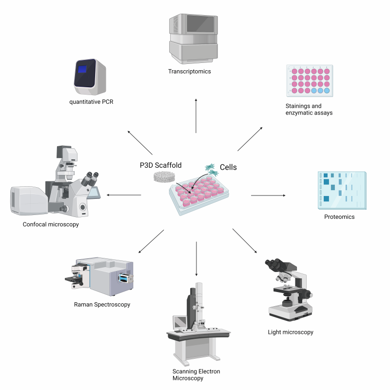
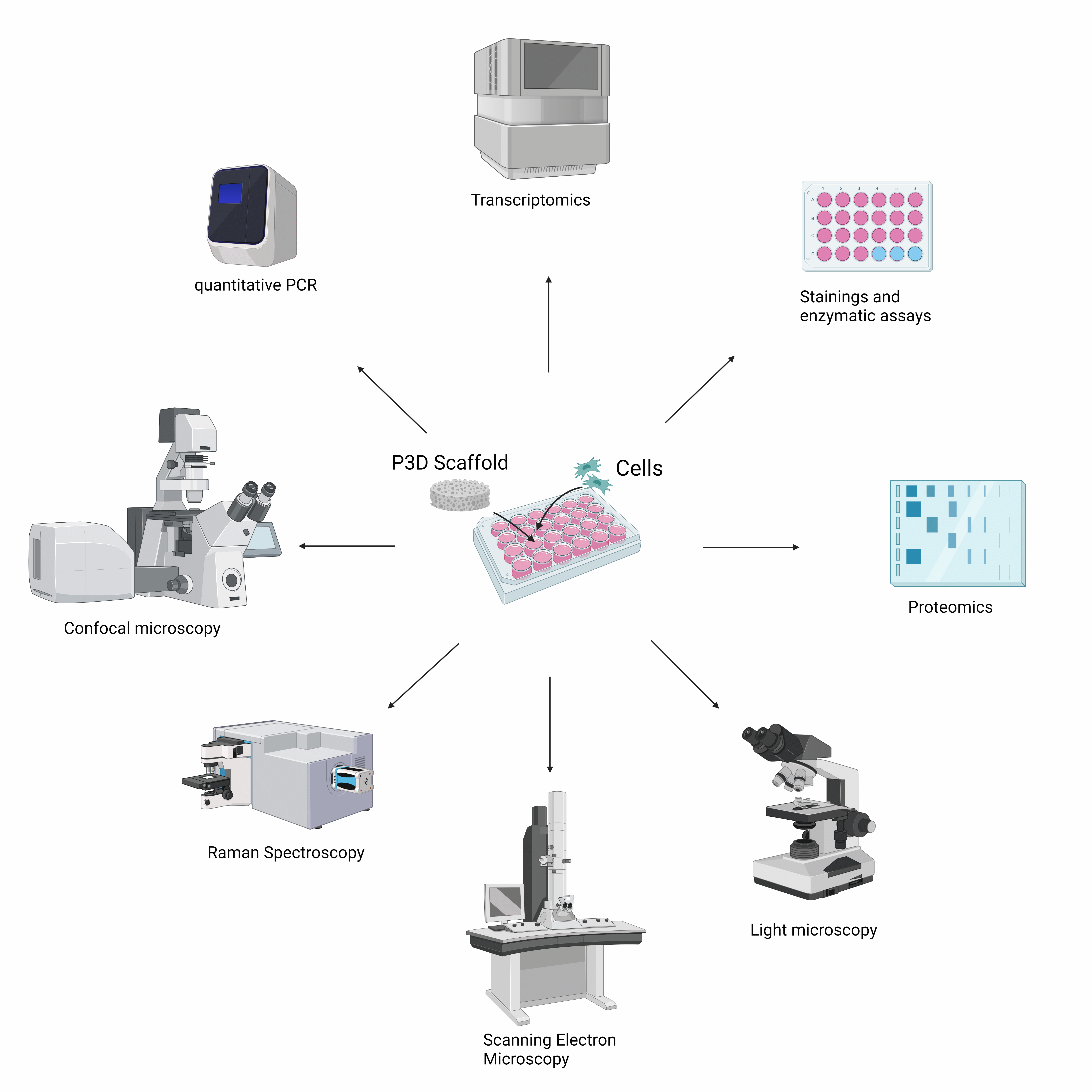
Figure 1. Overview of analytical techniques compatible with P3D Scaffolds, illustrating how structural, molecular, and functional analyses can be integrated within a single 3D culture system.
A common question when adopting a new 3D culture system is whether existing laboratory techniques can still be applied effectively. One of the key advantages of P3D Scaffolds is that they are designed to integrate seamlessly into standard laboratory workflows. This allows researchers to integrate the scaffolds into existing workflows without the need for specialized equipment or major protocol changes.
Most traditional laboratory techniques are compatible with the P3D Scaffolds, including:
Together, these techniques support the full experimental workflow, from initial cell seeding and culture to advanced downstream analyses. This flexibility allows researchers to combine structural, functional, and molecular insights within the same 3D system.
These capabilities enable a wide range of applications. For example, researchers can investigate cell–cell interactions in a more physiologically relevant environment, study tissue development and differentiation, or evaluate how biomaterials interact with living cells. In addition, the scaffolds support studies of microbial colonization and host–microbe interactions in three dimensions. The ability to combine structural analysis with molecular readouts makes P3D Scaffolds particularly well suited for translational and preclinical research.
Depending on where you are in your workflow, different resources may be most helpful. If you are just getting started, our Protocols page and Frequently Asked Questions page provide a solid foundation for the early stages of planning. For an overview of scaffold properties, recommended use, and experimental considerations, the Technical Datasheet provides essential background information, while our Publications provide validation through peer‑reviewed studies. If you prefer visual guidance, our Video Protocols provide step‑by‑step demonstrations.
As 3D cell culture models continue to evolve, the need for standardized and reproducible workflows becomes increasingly important. We view these materials as living resources that will continue to develop alongside new applications and insights from the research community.
By consolidating our technical knowledge, we aim to reduce the barrier to entry for 3D cell culture with P3D Scaffolds and to enable researchers to focus on generating meaningful biological insights. We look forward to seeing how the research community applies these tools and contributes to advancing 3D cell culture methodologies.
