")

New scientific paper on Ossiform’s P3D Bone implant
New scientific paper on Ossiform's P3D Bone
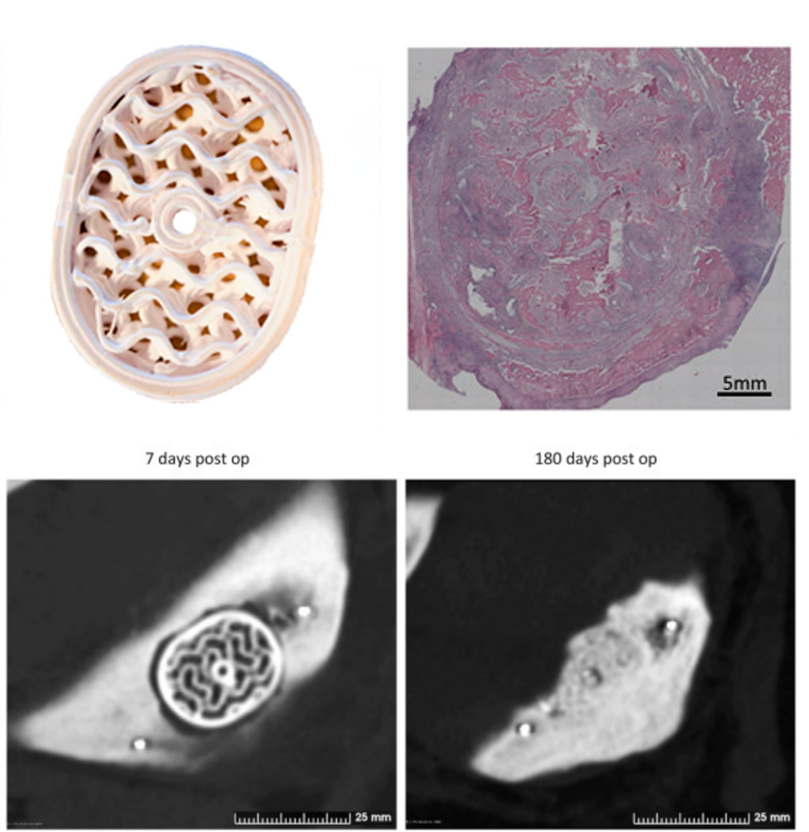
Ossiform has compared its 3D-printed P3D Bone implant with an off-the-shelf b-TCP implant. The implants were inserted in the mandible ramus of pigs. After six months, resorption and remodeling were evaluated using CT-scans and histological images.
After six months, the P3D Bone implants were almost completely resorbed and replaced by vascularized compact bone. This also allowed osteointegration to occur. The histological image in the upper right corner shows the partially resorbed P3D Bone implant after three months.
The results also showed that the off-the-shelf implant and the P3D Bone performed equally well. This includes the predicted osteointegration medially and laterally and minimal gaping between the implants and native bone.
The full results supported by both histology and CT data are published in Bone, Volume 159, June 2022.

Conduct your own in vivo studies using Ossiform Research Line
The P3D Bone graft substitute has not yet reached the market. However, Ossiform’s research product line enables you to perform implant studies with the company’s material and technology.
The P3D Scaffolds are made of the same material as the P3D Bone. They are 3D printed to cater to your research needs. They are approved for animal studies and previous studies demonstrate their usability in mice (Jensen et al, 2020).
The P3D Scaffolds are also suitable for in vitro research. Thereby opening a world of opportunities for you and your research. For instance, the P3D Scaffolds enable you to seed selected cell cultures on the scaffold before implantation. This makes them advantageous for cancer modelling and various other studies, where the gap between in vitro and in vivo research may seem unbridgeable.