​News from the Lab

Technical data sheet available for P3D Scaffolds

Biomimetic scaffolds

Technical data sheet available for P3D Scaffolds

This year, we have been busy in the lab fine tuning the small details and testing various methods and techniques in combination with the biomimetic 3D cell culture systems, P3D Scaffolds.

Now we can share that all the testing has been boiled down to this technical data sheet. Here you will find general guidelines, relevant flow charts, and links for all our protocols and other materials for researchers. Futhermore, you can access all of P3D Scaffolds technical specifications and recommended seeding densitites when adding cells.

Figure that shows a selection of standard analysis methods that are compatible with P3D Scaffolds, including light and confocal microscopy, SEM, transciptomics, raman spectroscopy ,and quantitative PCR

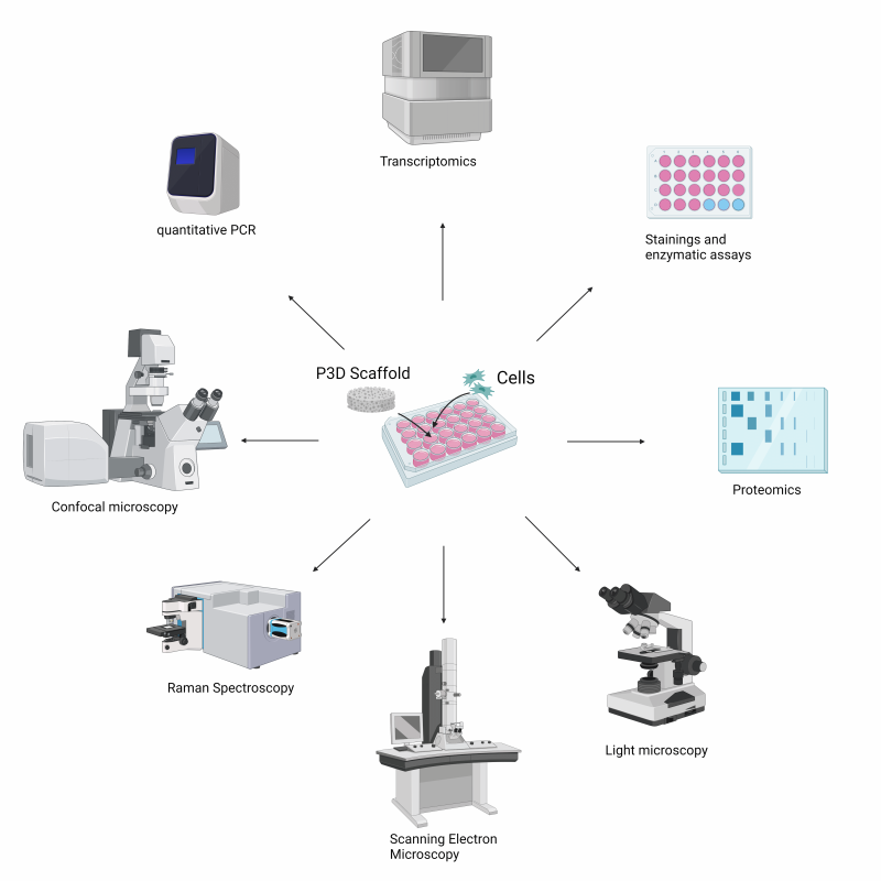
Most traditional laboratory techniques are compatible with the P3D Scaffolds, including:

  • Inverted light microscopy (through the pores)
  • Fluorescence microscopy such as for human or bacterial cells that are labelled with green fluorescent protein (GFP)
  • Nucleic acid extraction for PCR and next generation sequencing (NGS)
  • Staining assays such as an ALP staining assay. The dye may be detached from the 3D structures after staining for quantification using absorbance
  • Enzymatic assays such as the p-nitrophenyl phosphate based assay for alkaline phosphatase activity
  • Micro-computed tomography (microCT) imaging
  • Scanning electronic microscopy (SEM)
  • Energy-dispersive X-ray spectroscopy (EDX/EDS)
  • X-ray fluorescence (XRF)
  • Raman spectroscopy
  • Cell recovery by trypsinization (for single cell analyses/methods like FACS and single cell​sequencing)​
  • Confocal microscopy (LCSM)
  • CellTiter-Glo 3D cell viability assay
  • Live/Dead Stain
  • Western Blot

Explore all product materials, protocols, research handbook, and data sheets here

Watch all video protocols on Ossiform’s YouTube Channel here