Contact us here!
Contact us for questions, general inquiries, or to request a quote.
By submitting this form, I consent to the processing of my personal data as explained in Ossiform's Privacy Policy.
Contact us for questions, general inquiries, or to request a quote.
By submitting this form, I consent to the processing of my personal data as explained in Ossiform's Privacy Policy.
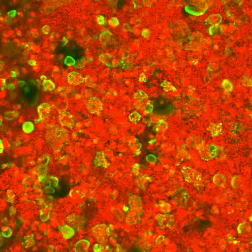
CD14+ monocytes were isolated from blood of human donors and subsequently differentiated into mature osteoclasts. The mature osteoclasts were cultured on rhodamine (red) stained P3D Scaffolds and f-actin (green) in living osteoclasts. This was labeled using SiR-actin.
A time-lapse was conducted over a period of 70 hours. The images was acquired every 21 minutes. By employing time-lapse microscopy, it was possible to confirm the osteoclast resorption of the scaffold.
Read the newest peer-reviewed scientific publication using P3D Scaffolds here.
In the study, microcomputed tomography was utilized to investigate the microstructural parameters of BTCP-based bone-mimicking scaffolds, the P3D Scaffolds. Here, high and low-resolution microCT was used to quantitatively evaluate the 3D-printed, bone-mimicking constructs in terms of surface area, construct volume, number of pores, pore size and distribution, wall thickness, delamination, internal porosity, and internal structure. In addition to establishing these parameters in Ossiform’s P3D Scaffolds, this article underlines the value of microCT as a tool in the quality management of bioceramic implants.